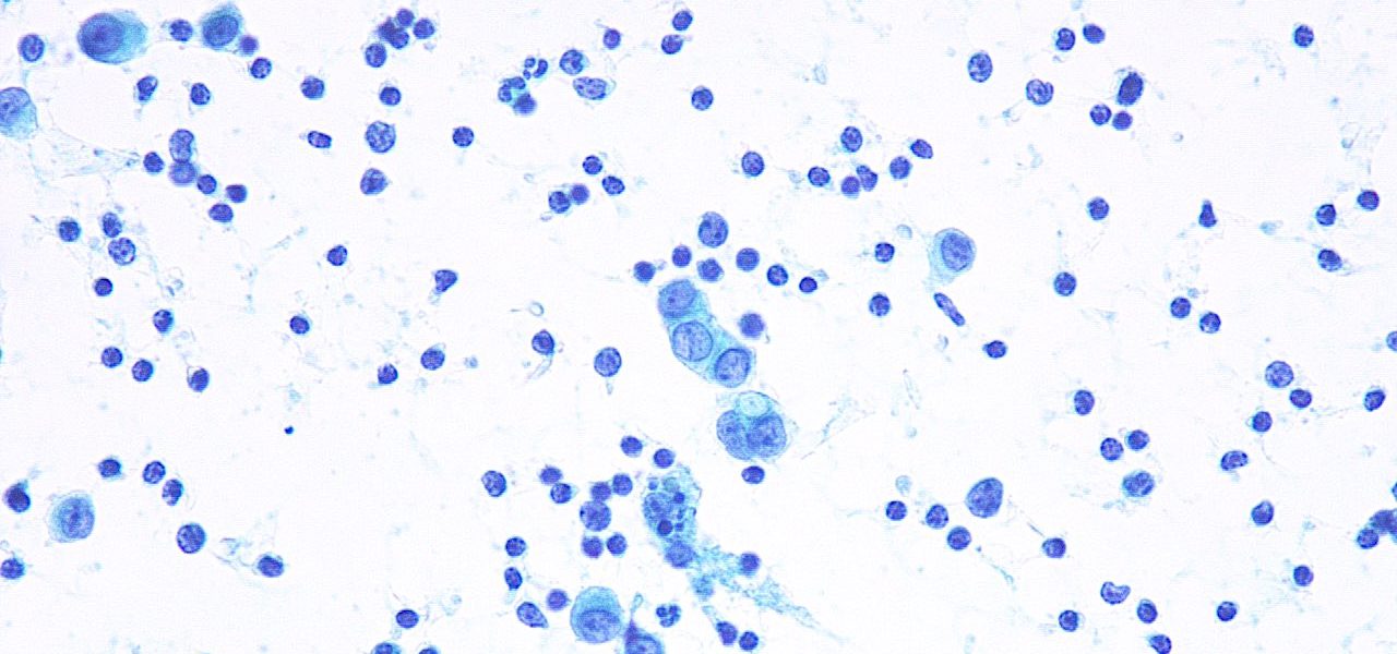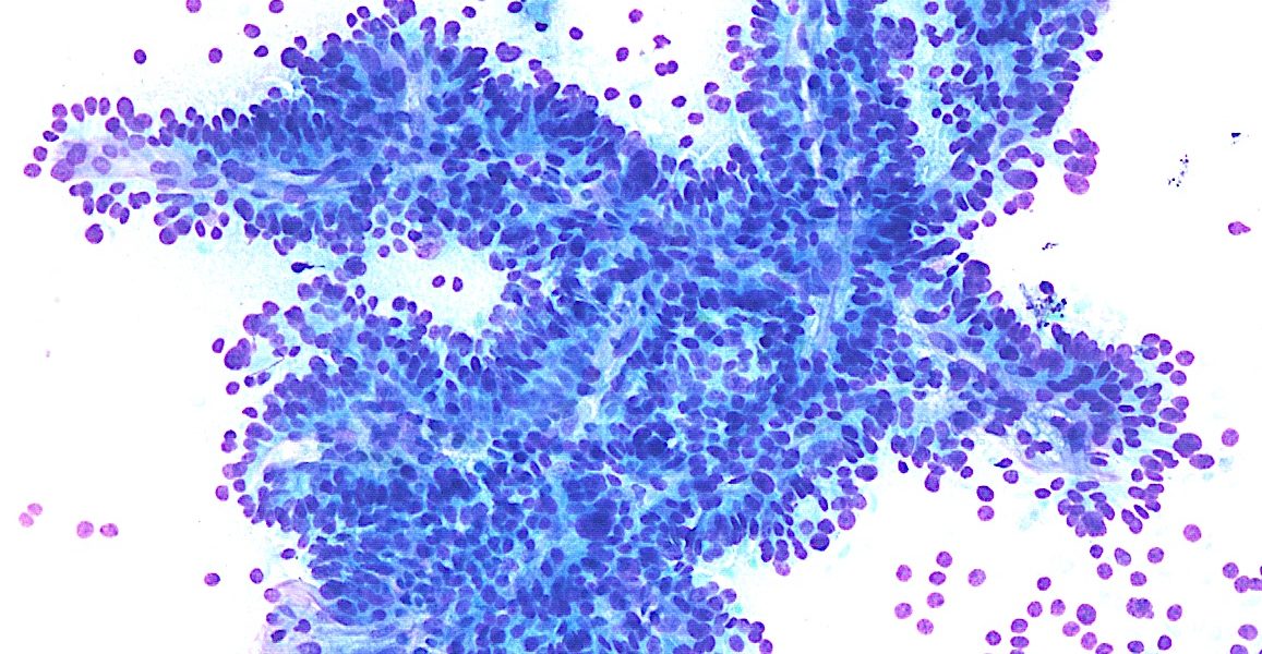
FNA of a clinically aggressive thyroid tumor showing morphologic features suggestive of columnar cell variant of papillary thyroid carcinoma. Image 2: Evidence of anaplastic transformation is seen in the following picture: Credit: Dr. Yazeed Alwealie
أحدث الفعاليات
- ديسمبر 2024
-
-
CSF: Metastatic lobular carcinoma Microscopic description: Glandular cells in “Indian-file” pattern, prominent nucleoli and targetoid cytoplasm Credit: Dr. Yazeed Alwelaie
-
انضم الى فريق النشاط التعليمي لدينا لتصبح متحدثا او صانع محتوى
-
تابع آخر أخبارنا و مقالاتنا وانشطتنا وفعاليتنا على تيليغرام
-
بمناسبة تدشين أعمال الجمعية السعودية لتقنيتي الخلايا و الانسجة ، فإن الجمعية تقدم عضويات مجانية لفترة محدودة يسعدنا انضمامكم لنا.
-
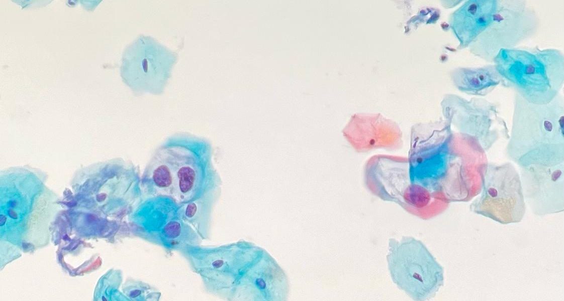
Microscopic descriptionAtypical squamous cells exhibiting enlarged nuclei (>3x normal intermediate squamous cells), irregular nuclear membrane contours, hyperchromasia and perinuclear halo (koilocytic changes). InterpretationLow grade Squamous Intraepithelial Lesion (LSIL) CreditAbdulaziz Assiri, MSc, CT(ASCPi)HTL Aisha Althenian, MSc, CT(ASCP)
- لاتوجد مواضيع اخرى لعرضها