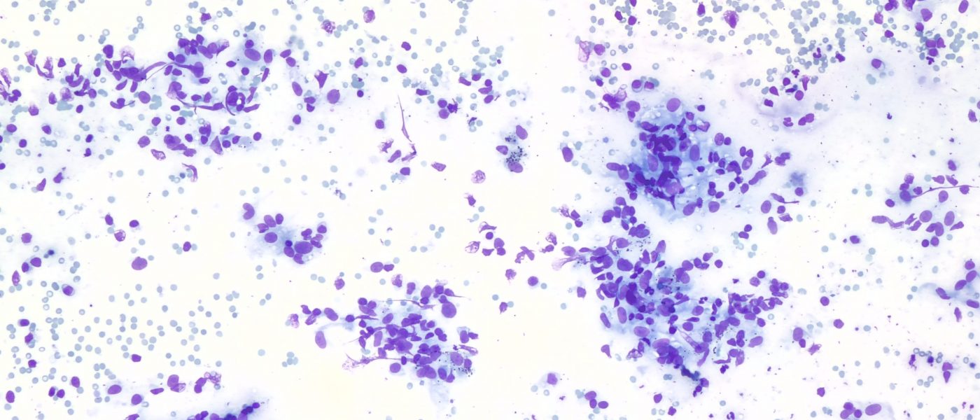
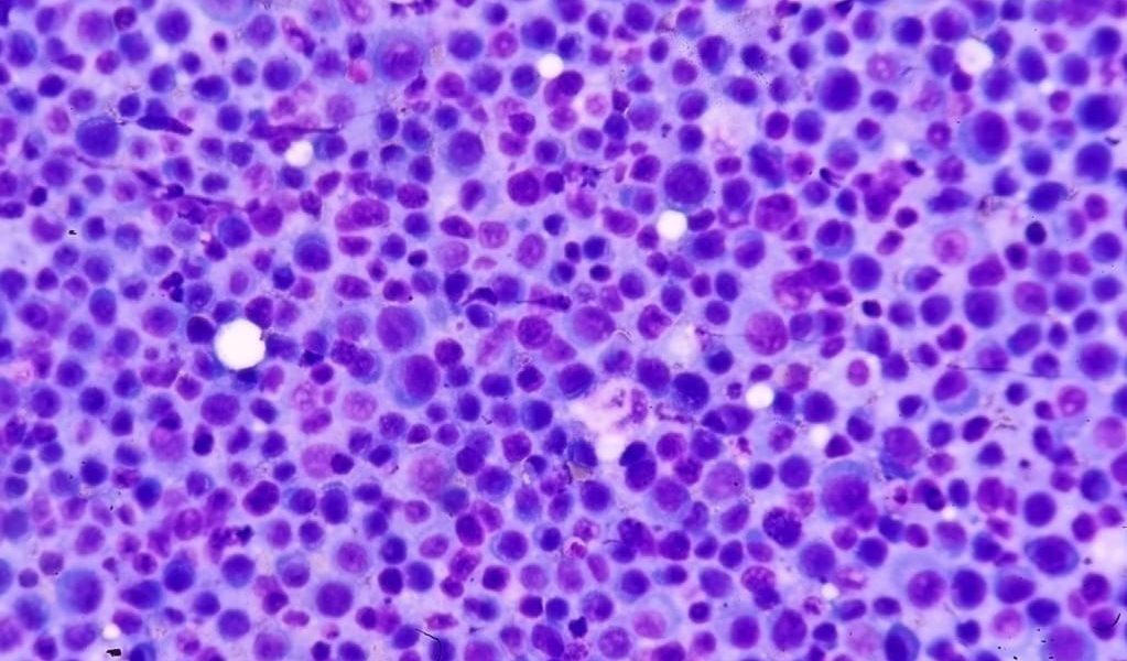
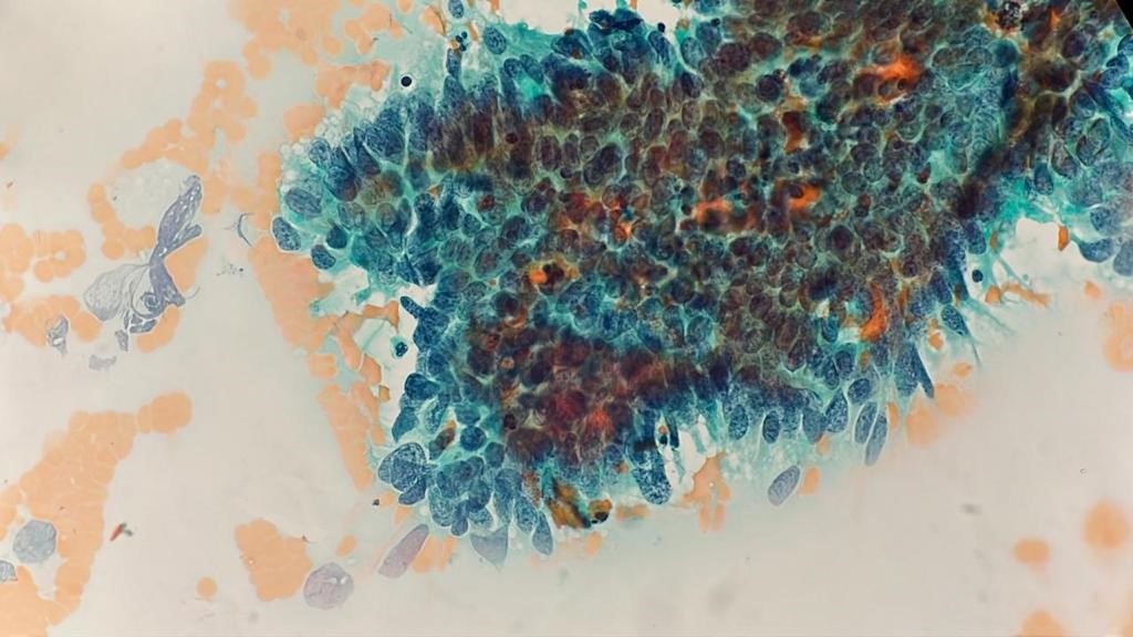
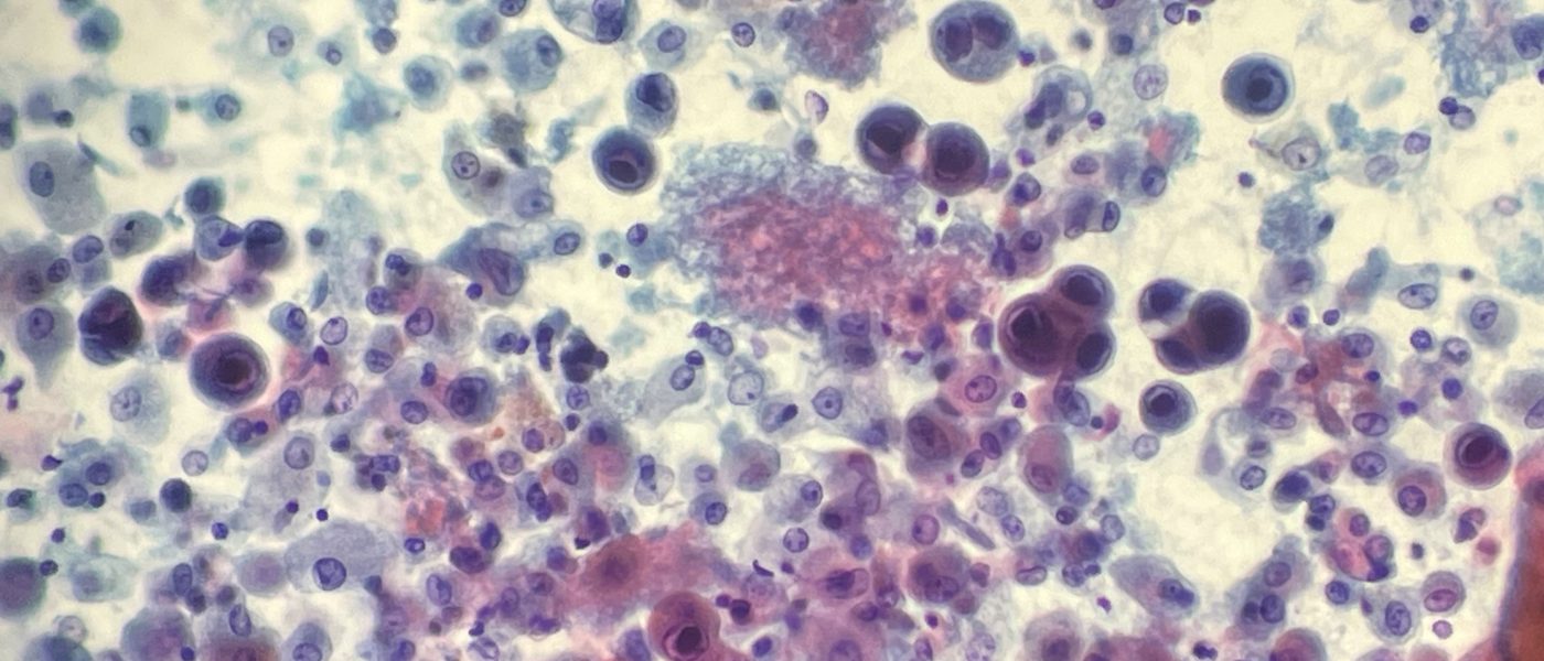
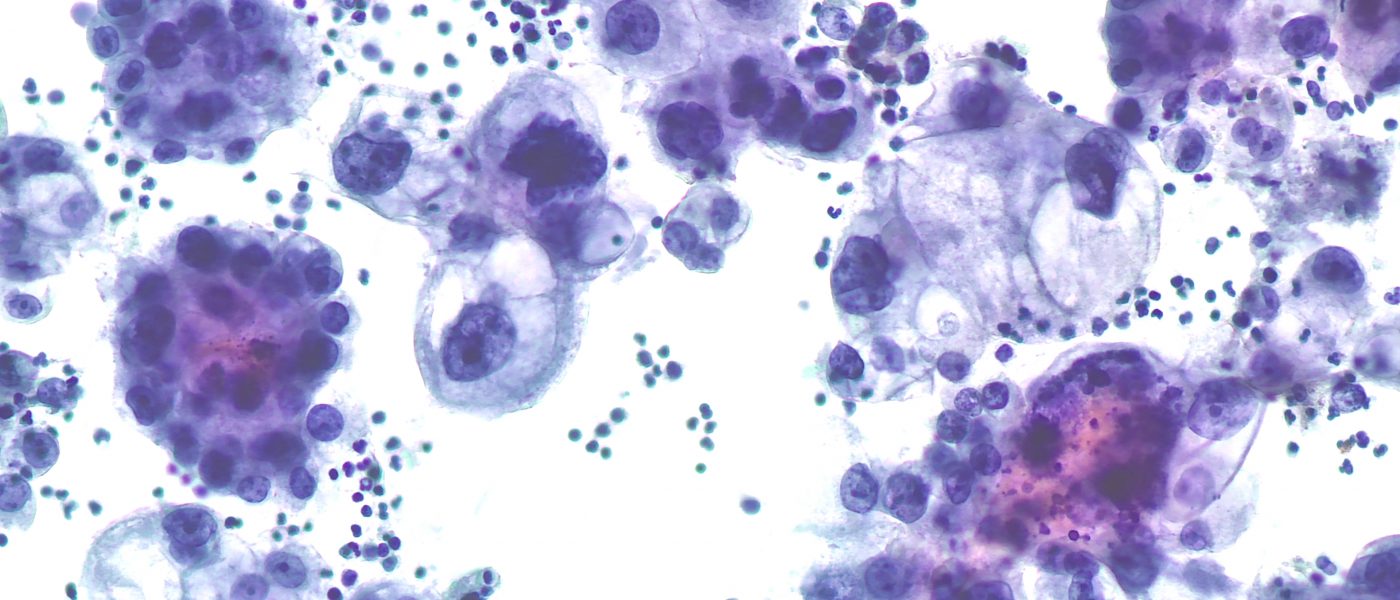
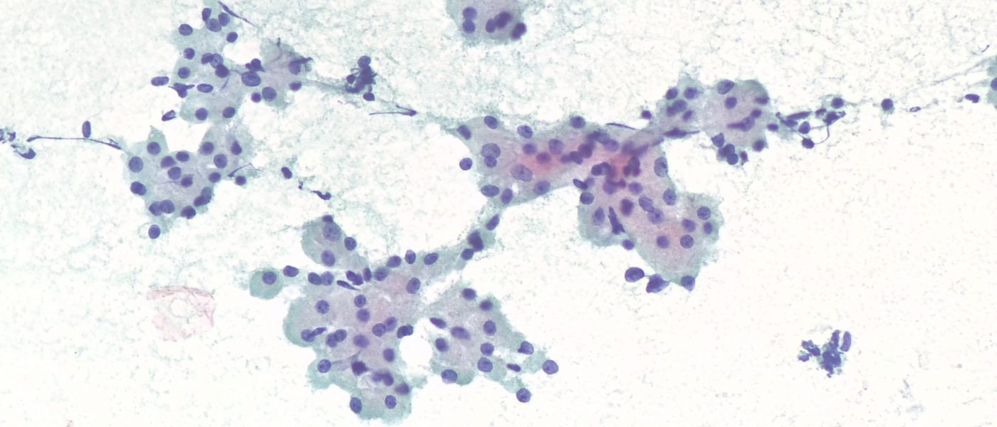

Hilar Lymph Node (EBUS-FNA) Diagnosis: Granulomatous lymphadenitis (non-necrotizing) Credit: Mr. Moammar M. Muzzaffar, MSc, CT(IAC), CT(ASCP)
He presented with T cord compression secondary to bone lesions. This is an aspiration of a praspinal mass Specimen Type Paraspinal FNA Diagnosis Myeloma Credit: Ahmed Alsayed MSc, CT(IAC), CT(ASCP), CPHQ
-Ascitic fluid- General Diagnostic Category: Malignant cells identified The International System for Reporting Serous Fluid Cytopathology, category: MAL-S (Malignant Secondary) Inerpretation: Metastatic breast lobular carcinoma Credit: Abdulellah Atia Alshareef, CT(IAC), CT(ASCP)
Clinical Information 39 years with abnormal bleeding Test Performed PAP SMEAR Conventional Specimen AdequacySatisfactory for evaluation; endocervical/transformation zone component present Interpretation (Abnormal)AIS Credit: Abdulellah Atia Alshareef, CT(IAC), CT(ASCP)
Test PerformedSurePath™ PAP SMEAR Specimen AdequacySatisfactory for evaluation; endocervical/transformation zone component present Interpretation (Abnormal)High-grade Squamous Intraepithelial Lesion (HSIL) Credit: Moammar M. Mudhaffar, MSc, CT(IAC), CT(ASCP)
-Bronchoalveolar Lavage- :General Diagnostic Category Negative for malignancy :Interpretation POSITIVE for Pneumocystis organisms- Numerous Cytomegalovirus inclusion identified- Credit: Mr. Moammar M. Muzzaffar, MSc, CT(IAC), CT(ASCP)
-Pleural effusion- General Diagnostic Category: Malignant cells identified The International System for Reporting Serous Fluid Cytopathology, category: MAL-S (Malignant Secondary) Inerpretation: Metastatic Breast Ductal Carcinoma Credit: Moammar Mudhaffar, MSc, CT(IAC), CT(ASCP)
-Thyroid FNA- General Diagnostic Category : Malignant (Category VI) Interpretation: Non-Hodgkin lymphoma ( High grade B-cell Lymphoma). :Microscopic description Cellular aspirates that show predominance of medium to large lymphocytes with abnormal cytologic features. Scattered follicular cells and Hurthle cells noted Flow Cytometry Summary: Immunophenotyping of Fine Needle Aspiration leukocytes by flow cytometry shows an abnormal […]