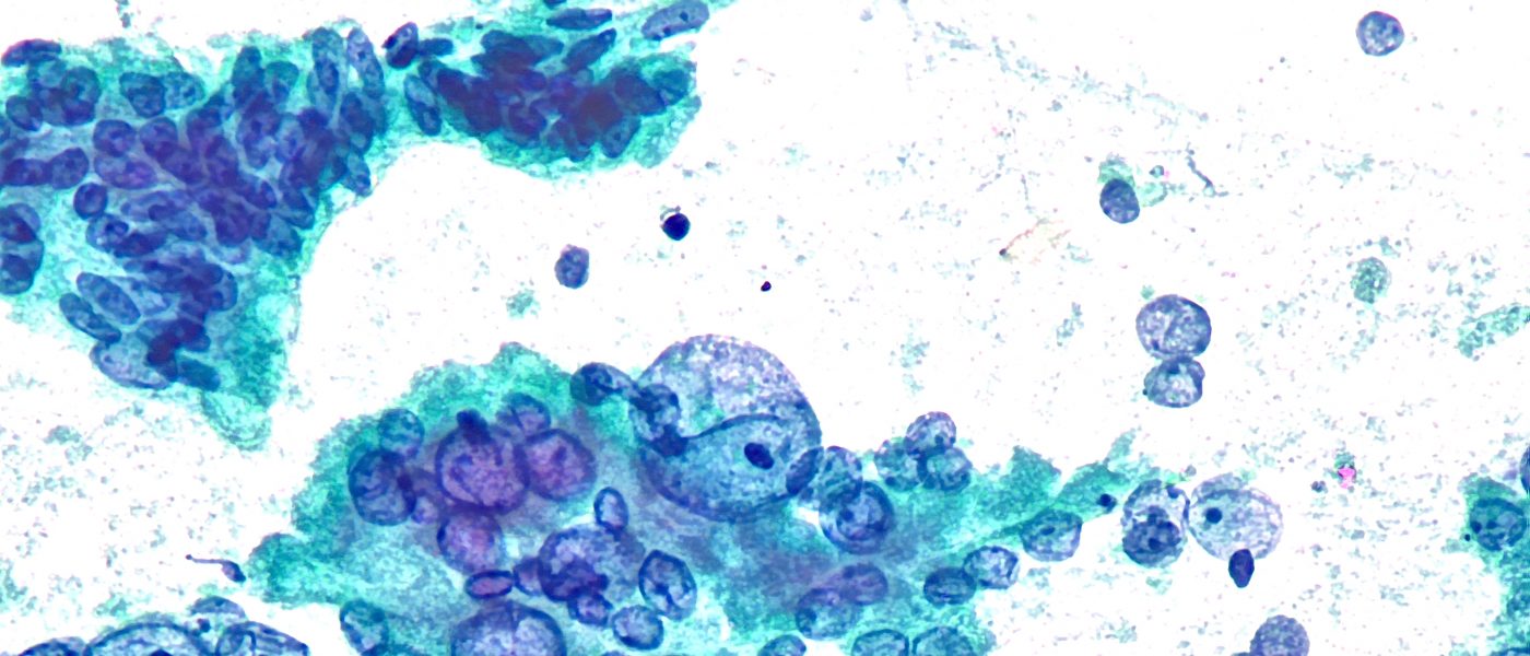
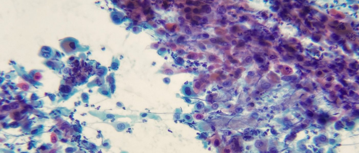
Cytologic features of pancreatic adenocarcinoma are depicted in this pap-stained smear. Marked size variability (anisonucleosis), nuclear membrane irregularity and pale/hypochromatic nuclei are helpful features. Credit: Dr. Yazeed AlWelaie
Cytologic features of pancreatic adenocarcinoma are depicted in this pap-stained smear. Marked size variability (anisonucleosis), nuclear membrane irregularity and pale/hypochromatic nuclei are helpful features. Credit: Dr. Yazeed AlWelaie
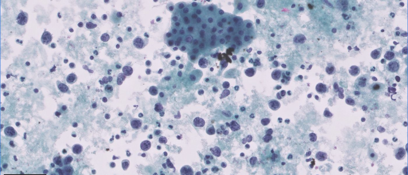
EUS-FNA of Lymphoepithelial Cyst of the Pancreas showing nucleated and anucleated squamous cells with scattered lymphocytes in the background. Credit: Dr. Yazeed Alwelaie
Liver FNA Diagnosis: B-cell Lymphoma, confirmed by IHC Credit: Abdulelah Attiah, CT(ASCP), CTIAC

FNA , Multiple Liver lesions . Metastatic adenocarcinoma Credit: Abdulelah Attiah, CT(ASCP), CTIAC
FNA , Liver mass Dx : Carcinoid tumor Credit: Abdulelah Attiah, CT(ASCP) CTIAC
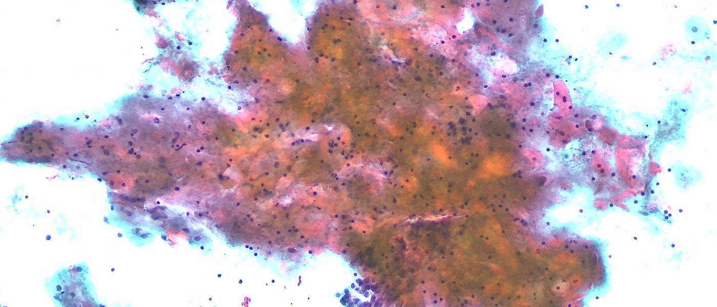
Pancreatic mass FNA showing poorly differentiated carcinoma with extensive squamous differentiation. Credit: Dr. Yazeed Alwelaie
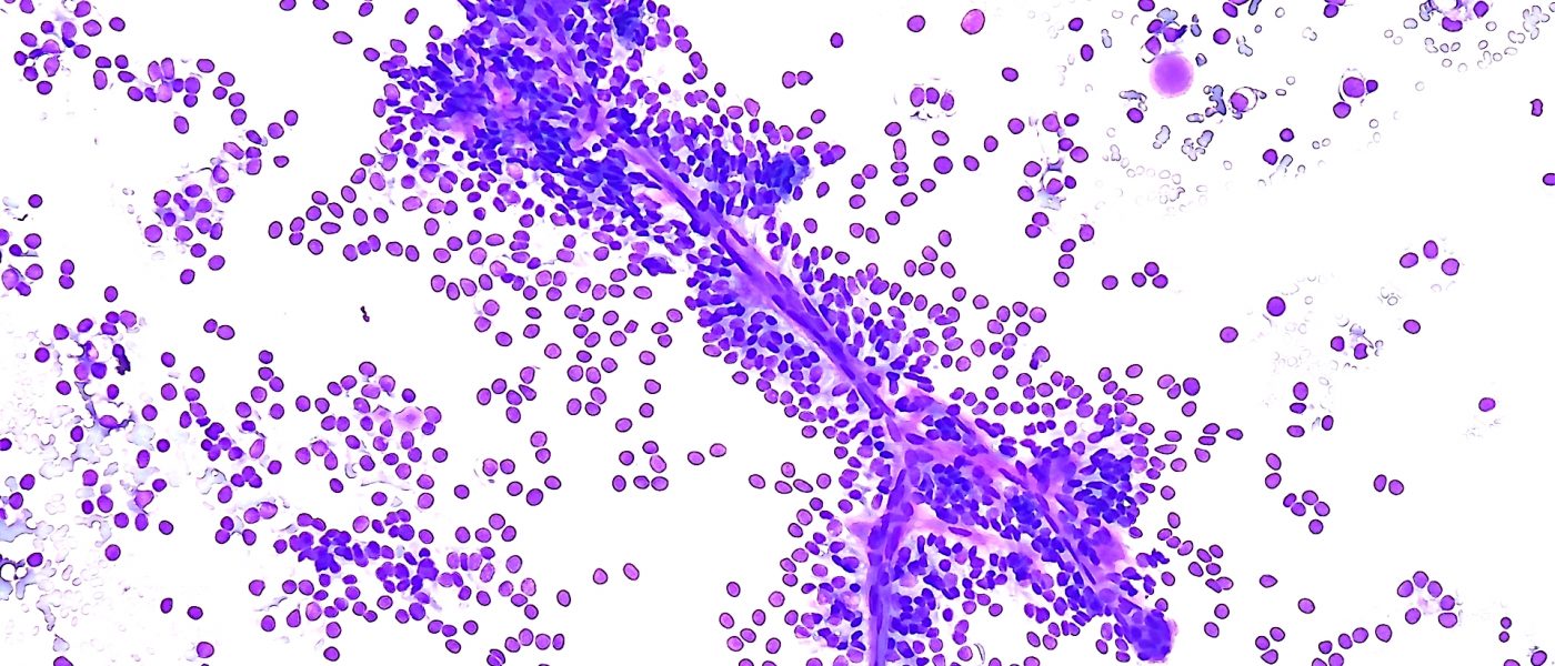
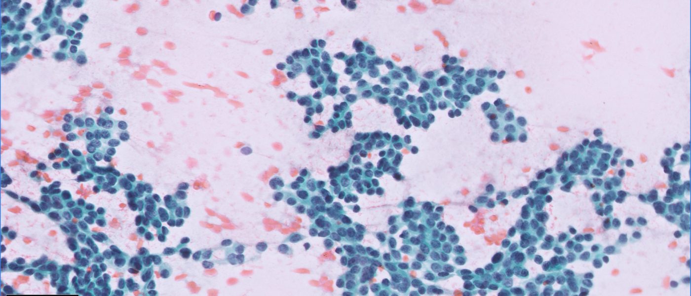
Solid pseudopapillary neoplasm (SPN) of the pancreas on direct cytologic smears obtained during on-site evaluation. Neoplastic cells are seen clinging to the branching delicate capillaries. Credit: Dr. Yazeed Alwelaie