Category Archives: Salivary gland FNA

  • Case #8

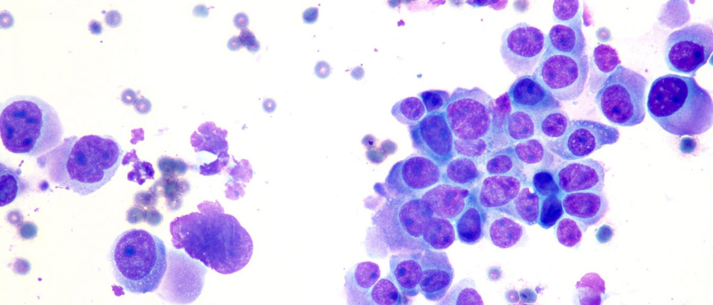
    General Diagnostic Category : Neoplasm: Benign (Category IV)  Interpretation: Cellular oncocytic/oncocytoid neoplasm Microscopic description : Smears show oncocytic cells in a background of blood and mixed inflammatory cells. Credit: Moammar M. Mudhaffar, MSc, CT(IAC), CT(ASCP)

  • Case 7

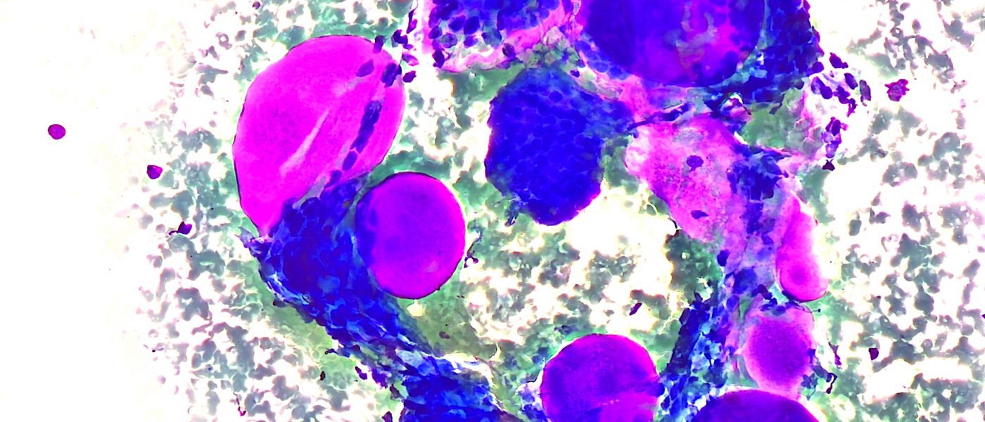
    Submandibular mass .Diff Quick smears and cell block . Diagnosis  : Adenoid cystic carcinoma ( ACC ) . The diagnostic clue in aspirates from ACC are large globules of extracellular matrix, partially surrounded by basaloid tumor cells. Credit: : Abdulelah Attiah , CT(ASCP),CTIAC 

  • Case 6

    Bubblegum-like, well-defined hyaline globules of adenoid cystic carcinoma on Diff-Quik smear from a neck mass fine needle aspiration. Credit: Dr. Yazeed Alwelaie

  • Case 5

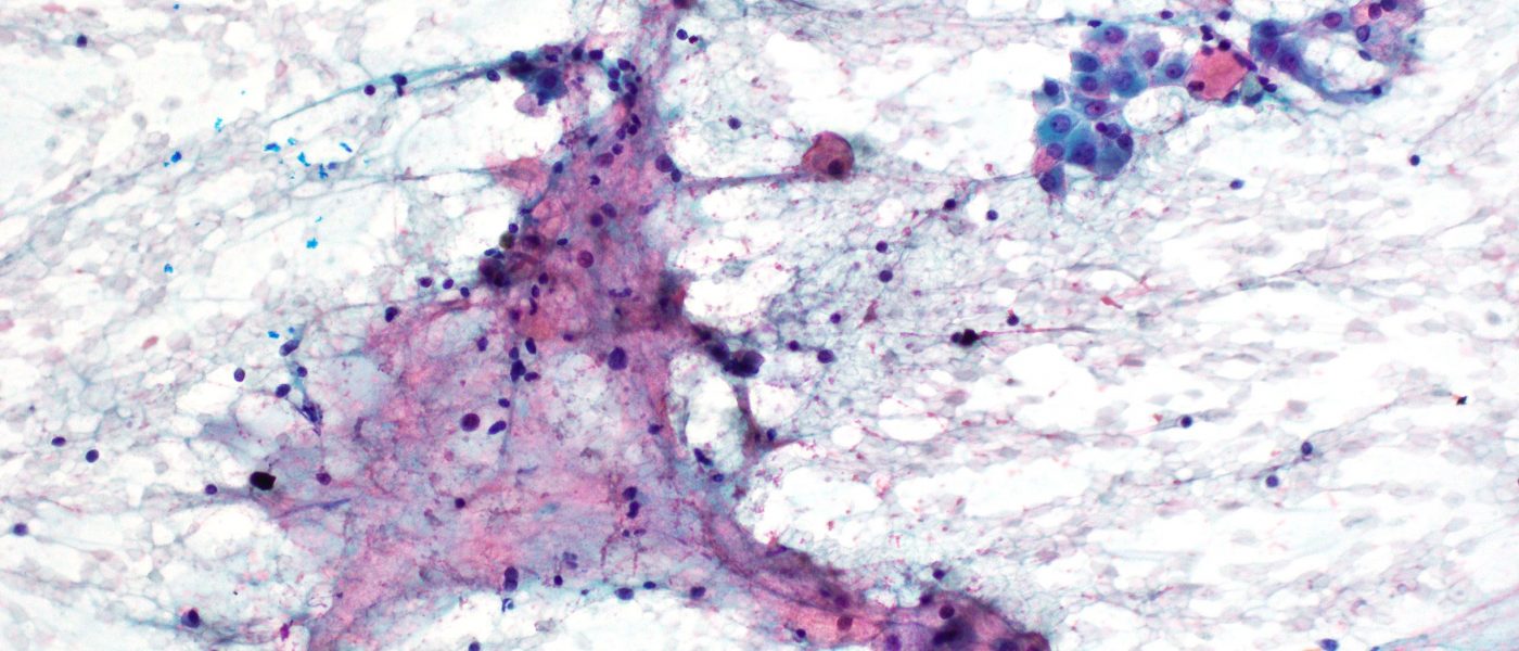
    #FNA of a small submandibular mass showing a nice example of mucoepidermoid carcinoma. Smears show a combination of mucous and intermediate cell associated with mucin in the background. The inflammatory component is a response to extravasated mucin. Credit: Dr. Yazeed Alwelaie

  • Case 4

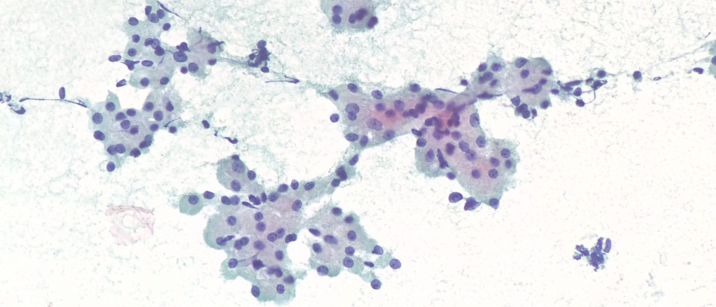
    #FNA of a parotid mass showing ACINIC CELL CARCINOMA – large acinar sheets that lack ducts and with finely granular to vacuolated cytoplasm. Credit: Dr. Yazeed Alwelaie

  • Case 1

    FNA of a rapidly enlarging parotid mass showing classic cytomorphology of salivary duct carcinoma (SDC) with strong androgen receptor reactivity. Credit: Dr. Yazeed Alwelaie