
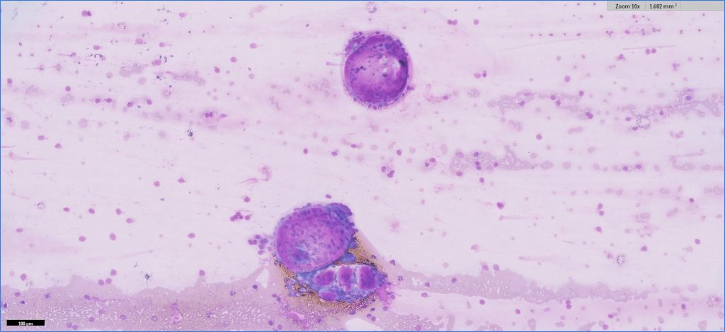
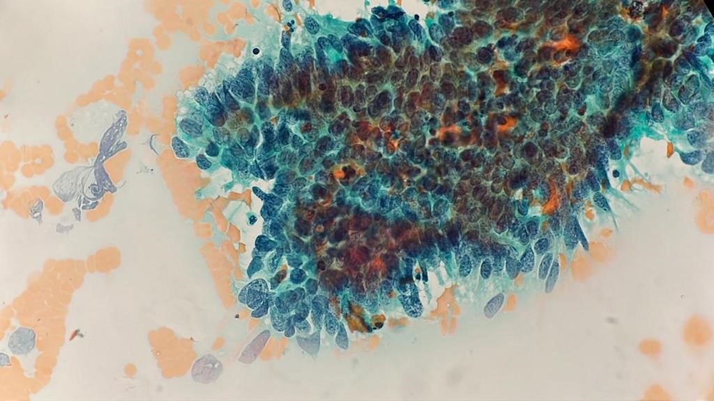
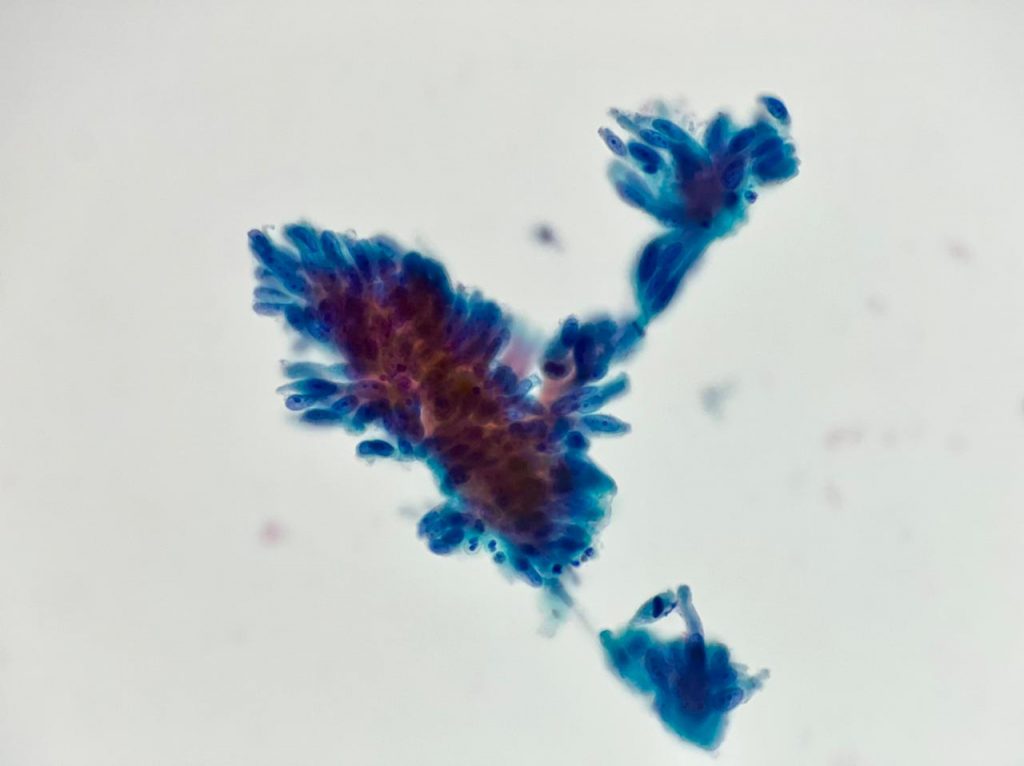
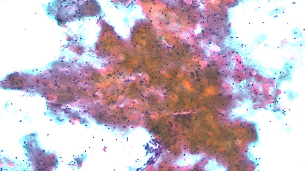
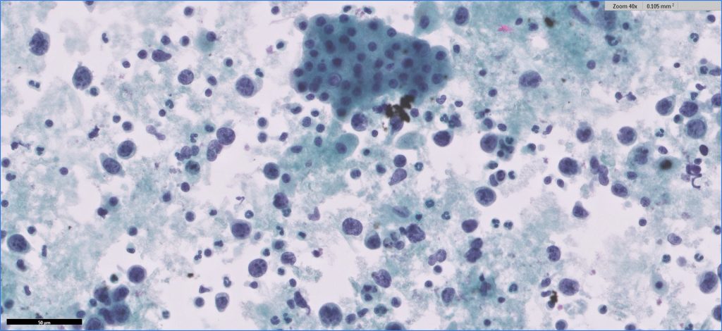
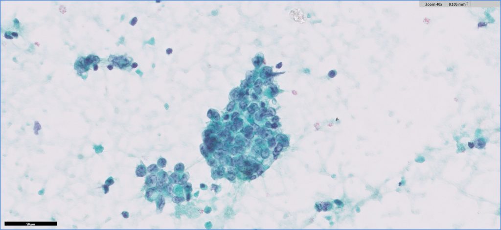
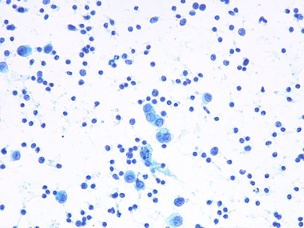
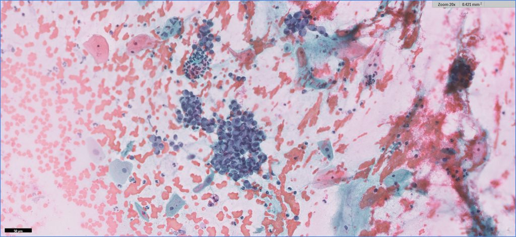
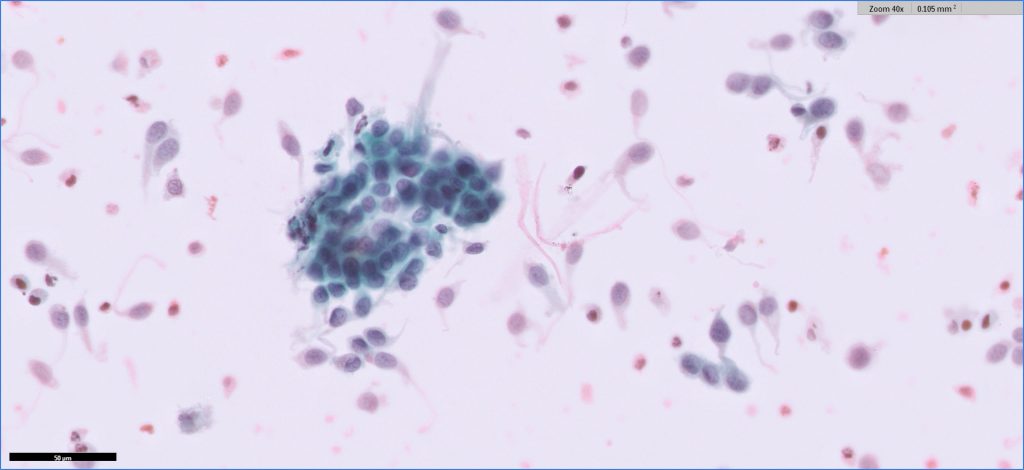
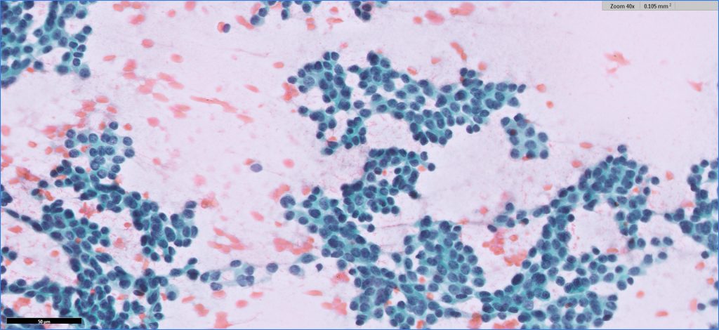
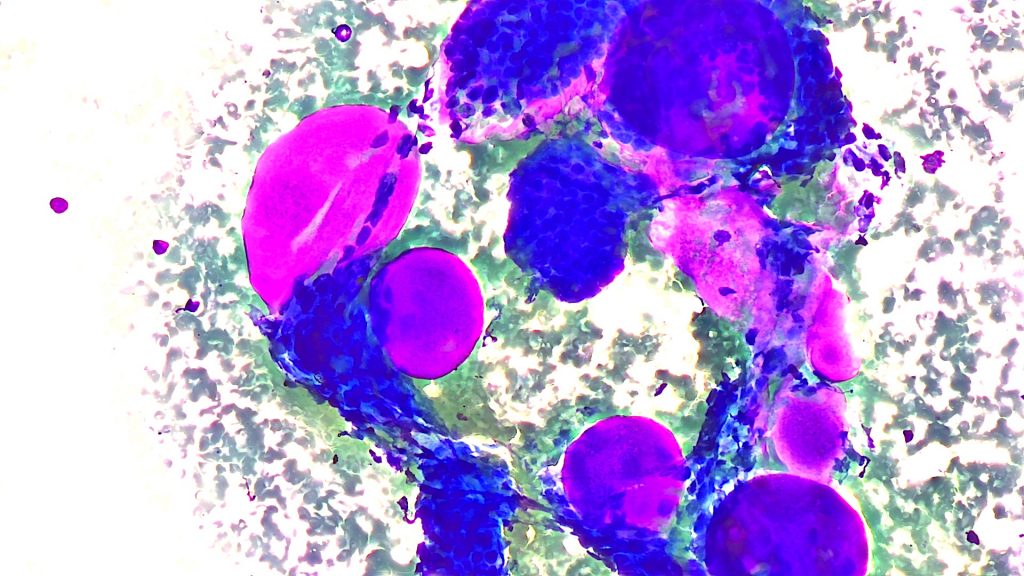
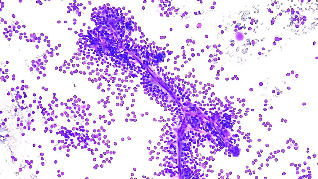
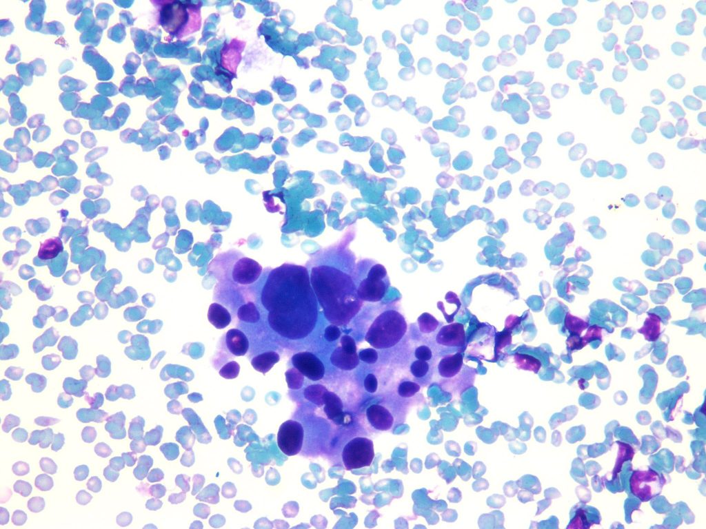
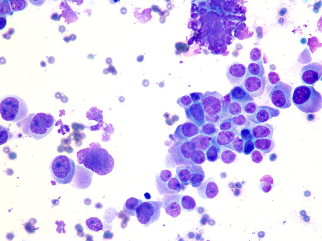
Case #8
General Diagnostic Category : Neoplasm: Benign (Category IV) Interpretation: Cellular oncocytic/oncocytoid neoplasm Microscopic description : Smears show oncocytic cells in a background of blood and mixed inflammatory cells. Credit: Moammar M. Mudhaffar, MSc, CT(IAC), CT(ASCP)
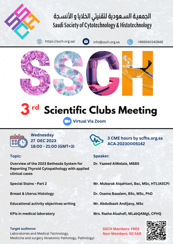
3rd Scientific clubs meeting of the SSCH
As we approach the end of the year, The Saudi Society of Cytotechnology and Histotechnology is delighted to invite you to the 3rd Scientific Clubs Meeting for the year 2023. Riveting topics by experienced professionals awaiting you. See you all there. https://docs.google.com/forms/d/e/1FAIpQLScoEXomhTN5N7t4a8yvvV34oODmIj_R02XUVUsKYIFmrd4ekA/viewform

Call for abstract: poster presentation
The scientific committee of the APHCC is delighted to call for abstracts: poster presentation. The deadline is 30th of Jan, 2024. We look forward to seeing your posters in the APHCC. Please scan the code or use the following link: https://aphcc-ksa.com/call-for-abstract

Anatomic Pathology and Histo-Cytotechnology Conference (APHCC) 2024
It is our great pleasure to cordially invite you to the 1st international conference arranged by the Saudi Society of Cytotechnology and Histotechnology & the Saudi Society of Anatomical Pathology. We welcome you to the Anatomic Pathology and Histo-Cytotechnology Conference (APHCC) 5th – 7th March, 2024 Voco Hotel, Riyadh Saudi Arabia aphcc-ksa.com

نظام العضويات الجديد
للدخول على نظام العضويات على موقع الجمعية السعودية لتقنيتيّ الخلايا والأنسجة ، يرجى الضغط على الرابط التالي واختيار نوع العضوية ثم الضغط على أيقونة التسجيل: https://member.ssch.org.sa/
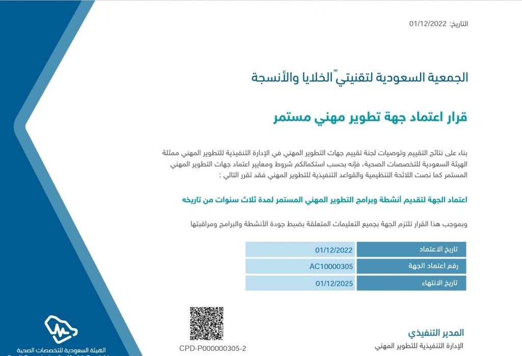
قرار اعتماد جهة تطوير مهني مستمر
تم بحمدالله اعتماد الجمعية السعودية لتقنيتيّ الخلايا والأنسجة كجهة مقدمة لأنشطة وبرامج التطوير المهني المستمر
Updates in Histotechnology‐part 1: 19 Mar 2023
Welcome to the 2nd educational activity of the Saudi Society of Cytotechnology and Histotechnology in 2023 Title: Updates in Histotechnology‐part 1: Principle of fixation, specimen processing, embedding, cutting and troubleshooting Date & Time: 19 Mar 2023, 16:00 – 21:00 CME: Accredited by the SCFHS for 3 CME hours Fees: 100 SAR SSCH members (Expired membership […]
Case 7
Submandibular mass .Diff Quick smears and cell block . Diagnosis : Adenoid cystic carcinoma ( ACC ) . The diagnostic clue in aspirates from ACC are large globules of extracellular matrix, partially surrounded by basaloid tumor cells. Credit: : Abdulelah Attiah , CT(ASCP),CTIAC
Case 5
Is there such a thing as pure signet ring cell carcinoma of lung? This is an EBUS-FNA of a hilar lymph node showing abundant neoplastic mucin admixed with loosely cohesive tumor cells. No history of primary tumor elsewhere. Credit: Dr. Yazeed AlWelaie

Case #2
-Bronchoalveolar Lavage- :General Diagnostic Category Negative for malignancy :Interpretation POSITIVE for Pneumocystis organisms- Numerous Cytomegalovirus inclusion identified- Credit: Mr. Moammar M. Muzzaffar, MSc, CT(IAC), CT(ASCP)
CASE # 2
-Ascitic fluid- General Diagnostic Category: Malignant cells identified The International System for Reporting Serous Fluid Cytopathology, category: MAL-S (Malignant Secondary) Inerpretation: Metastatic breast lobular carcinoma Credit: Abdulellah Atia Alshareef, CT(IAC), CT(ASCP)
Case 4
Routine Pap smear . Lower uterine segments (LUS ) , showing both epithelial and stromal components. Credit: Abdulelah Attiah, CT(ASCP), CTIAC
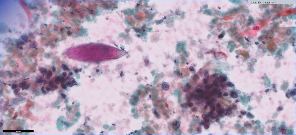
Case 1
Voided urine from a 59 years male with gross hematuria . Dx : Squamous cell carcinoma with Schistosoma ova Credit: Abdulelah Attiah , CT(ASCP), CTIAC

Laboratory safety #4
https://www.linkedin.com/feed/update/urn:li:activity:6996132873317621760

Case # 3
Hilar Lymph Node (EBUS-FNA) Diagnosis: Granulomatous lymphadenitis (non-necrotizing) Credit: Mr. Moammar M. Muzzaffar, MSc, CT(IAC), CT(ASCP)

Case # 2
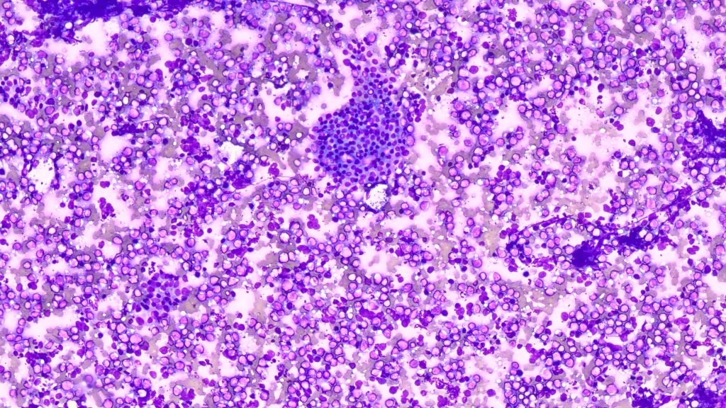
He presented with T cord compression secondary to bone lesions. This is an aspiration of a praspinal mass Specimen Type Paraspinal FNA Diagnosis Myeloma Credit: Ahmed Alsayed MSc, CT(IAC), CT(ASCP), CPHQ
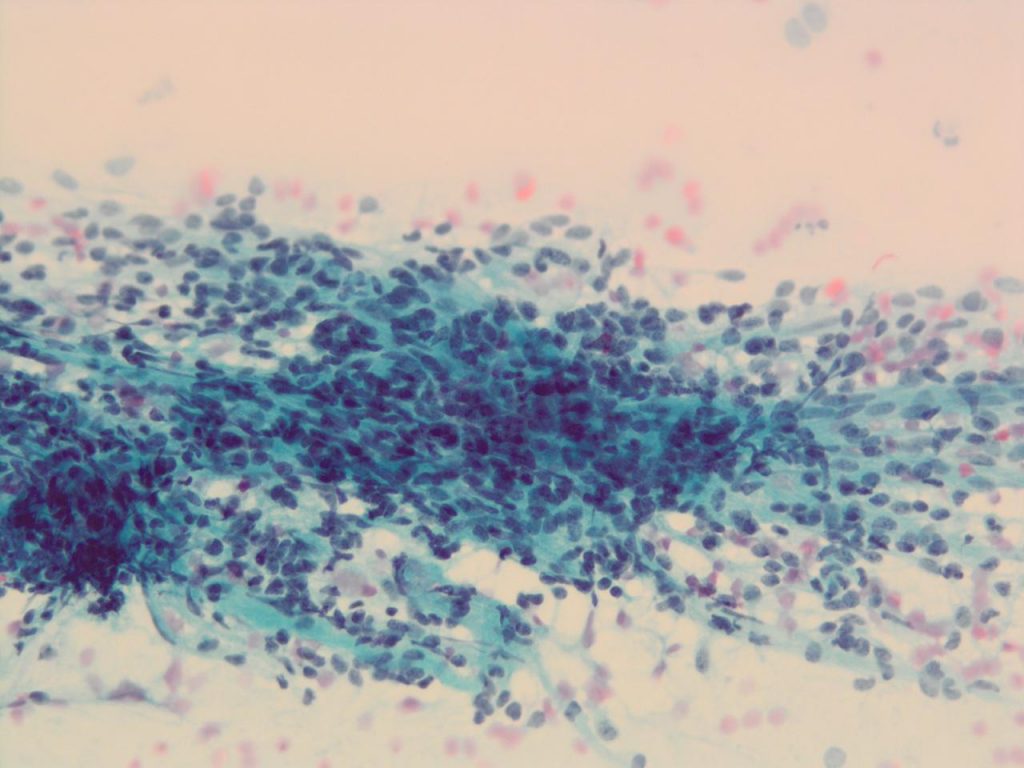
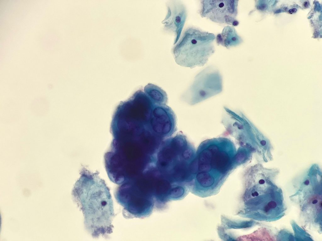
CASE # 8
Clinical Information 39 years with abnormal bleeding Test Performed PAP SMEAR Conventional Specimen AdequacySatisfactory for evaluation; endocervical/transformation zone component present Interpretation (Abnormal)AIS Credit: Abdulellah Atia Alshareef, CT(IAC), CT(ASCP)

Case # 7
Test PerformedSurePath™ PAP SMEAR Specimen AdequacySatisfactory for evaluation; endocervical/transformation zone component present Interpretation (Abnormal)High-grade Squamous Intraepithelial Lesion (HSIL) Credit: Moammar M. Mudhaffar, MSc, CT(IAC), CT(ASCP)
CASE #1
-Pleural effusion- General Diagnostic Category: Malignant cells identified The International System for Reporting Serous Fluid Cytopathology, category: MAL-S (Malignant Secondary) Inerpretation: Metastatic Breast Ductal Carcinoma Credit: Moammar Mudhaffar, MSc, CT(IAC), CT(ASCP)
1st Scientific clubs meeting 21 Mar 2023
For Registration: https://us06web.zoom.us/meeting/register/tZUofu-gqzIrHNEEWcgy6RrgJohxYPvfUyMk
Case # 6
-Thyroid FNA- General Diagnostic Category : Malignant (Category VI) Interpretation: Non-Hodgkin lymphoma ( High grade B-cell Lymphoma). :Microscopic description Cellular aspirates that show predominance of medium to large lymphocytes with abnormal cytologic features. Scattered follicular cells and Hurthle cells noted Flow Cytometry Summary: Immunophenotyping of Fine Needle Aspiration leukocytes by flow cytometry shows an abnormal […]

محضر اجتماع الجمعية العمومية للربع السنوي الأول لعام 2021
محضر-اجتماع-الجمعية-العمومية-للربع-السنوي-الأولتنزيل
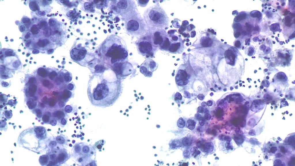
Case #6
Pap smear: – Cellular Changes Consistent with Herpes Simplex – Large multinucleated epithelial cells with 3Ms; Multinucleation, Molding, and Margination of chromatin. – Nuclei have a “ground-glass” appearance Credit: Abdulaziz Asiri, MSc, CT(ASCP), HTL(ASCPi)
Laboratory safety #6
https://www.linkedin.com/feed/update/urn:li:activity:7000086513879285760


Case 7
Cytologic features of pancreatic adenocarcinoma are depicted in this pap-stained smear. Marked size variability (anisonucleosis), nuclear membrane irregularity and pale/hypochromatic nuclei are helpful features. Credit: Dr. Yazeed AlWelaie
Case 5
Endocervical Adenocarcinoma – Abundant abnormal cells, typically with columnar configuration. – Enlarged, pleomorphic nuclei demonstrate irregular chromatin distribution, chromatin clearing, and nuclear membrane irregularities. – Macronucleoli. – Cytoplasm is finely vacuolated. Credit: Abdulaziz Asiri, MSc, CT(ASCP), HTL(ASCPi)
Case 6
EUS-FNA of Lymphoepithelial Cyst of the Pancreas showing nucleated and anucleated squamous cells with scattered lymphocytes in the background. Credit: Dr. Yazeed Alwelaie
Case5
Liver FNA Diagnosis: B-cell Lymphoma, confirmed by IHC Credit: Abdulelah Attiah, CT(ASCP), CTIAC


Free membership for a limited time
On the occasion of the inauguration of SSCH activities, memberships will be free for a limited period of time.


Case 2
CSF . 5 years male with pineal tumor . Dx : Pineoblastoma Credit: Abdulelah Attiah, CT(ASCP), CTIAC

Case 1
The type of cells that should never be in the CSF – history of gastric ca. Credit: Dr. Yazeed Alwelaie
Case 4
Lymph node: Metastatic lobular carcinoma Microscopic description: Glandular cells in “Indian-file” pattern, prominent nucleoli and targetoid cytoplasm Credit: Dr. Yazeed Alwelaie

Case 2
Voided urine from a patient with hematuria . 3D clusters with high N/C ratio , nuclear overlapping and fibrovascular core . Dx : Low grade urothelial carcinoma Credit: Abdulelah Attiah CT(ASCP),CTIAC
Case 3
Pap smear from a 53 patient with a known history of gastric cancer Microscopic Description: Loosely cohesive cluster and single atypical cells exhibiting hyperchromatic eccentric crescent shape nuclei ,coarse chromatin, irregular contour ,prominent nucleoli and vacuolated cytoplasm. Credit: Abdulelah Attiah , CT(ASCP),CTIAC
Case 1
FNA of neck mass from a 63 years male with history of bladder cancer . Dx : Metastatic urothelial carcinoma Credit: Abdulelah Attiah, CT(ASCP), CTIAC

Case 4
FNA , Multiple Liver lesions . Metastatic adenocarcinoma Credit: Abdulelah Attiah, CT(ASCP), CTIAC

Case 5
“Naked” papillary fronds in a thyroid FNA. Classic PTC cytomorpohlogy was seen throughout the smears. Credit: Dr. Yazeed Alwelaie
Case 6
Bubblegum-like, well-defined hyaline globules of adenoid cystic carcinoma on Diff-Quik smear from a neck mass fine needle aspiration. Credit: Dr. Yazeed Alwelaie
Case 2
Pancreatic mass FNA showing poorly differentiated carcinoma with extensive squamous differentiation. Credit: Dr. Yazeed Alwelaie
Case 5
#FNA of a small submandibular mass showing a nice example of mucoepidermoid carcinoma. Smears show a combination of mucous and intermediate cell associated with mucin in the background. The inflammatory component is a response to extravasated mucin. Credit: Dr. Yazeed Alwelaie
Case 4
#FNA of a parotid mass showing ACINIC CELL CARCINOMA – large acinar sheets that lack ducts and with finely granular to vacuolated cytoplasm. Credit: Dr. Yazeed Alwelaie
Case 1
Solid pseudopapillary neoplasm (SPN) of the pancreas on direct cytologic smears obtained during on-site evaluation. Neoplastic cells are seen clinging to the branching delicate capillaries. Credit: Dr. Yazeed Alwelaie
Case 3
Beautiful floret-like tyrosine crystals in a pleomorphic adenoma. Credit: Dr. Yazeed Alwelaie
Case 2
A nice #CytoPath example of ACINIC CELL CARCINOMA. Long-standing parotid mass. Cerdit: Dr. Yazeed Alwelaie

Case 2
#EBUS FNA of a mediastinal lymph node showing metastatic small cell carcinoma. Credit: Dr. Yazeed alwelaie
Case 1
FNA from an enlarged cervical lymph node showing a nice example of Hodgkin Reed-Sternberg cells. Credit: Dr. Yazeed Alwealie

Case 3
Nasopharyngeal carcinoma metastatic to cervical lymph node diagnosed on FNA. EBER ish and keratin + Credit: Dr. Yazeed Alwelaie
Case 4
Follicular neoplasm (oncocytic type) with prominent nuclear size variability. Credit: Dr. Yazeed Alwelaie
Case 1
FNA of a rapidly enlarging parotid mass showing classic cytomorphology of salivary duct carcinoma (SDC) with strong androgen receptor reactivity. Credit: Dr. Yazeed Alwelaie

Case 3
FNA of a thyroid lesion showing prominent globules reminiscent of adenoid-cystic carcinoma. High proliferative index in cellblock. Follow up: POORLY DIFFERENTIATED THYROID CARCINOMA. Credit: Dr. Yazeed Alwealie

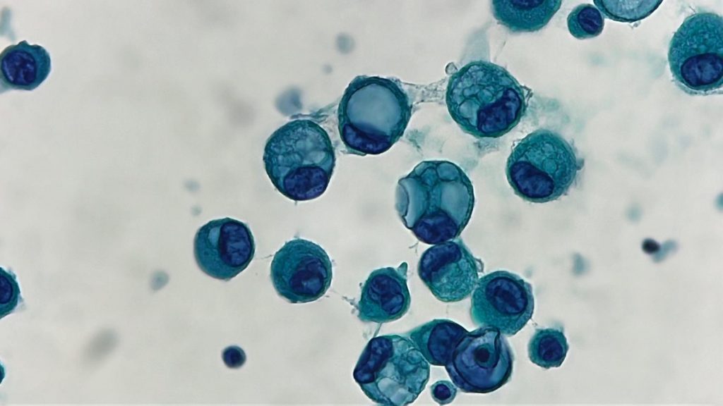
Case 1
Bronchoalveolar lavage showing numerous pneumocystis organisms and cytomegalovirus inclusions in a severely immunocompromised patient. Credit Dr. Yazeed Alwelaie Silver stain:
Case 2
Thyroid fine needle aspiration showing spindle cell cytomorphology of medullary thyroid carcinoma (MTC). Credit Dr. Yazeed Alwelaie
Case 2
Sneaky! There were many other similar cells in this SurePath pap. High-grade squamous intraepithelial lesion (HSIL). Credit: Dr. Yazeed Alwelaie

Case 1
FNA of a clinically aggressive thyroid tumor showing morphologic features suggestive of columnar cell variant of papillary thyroid carcinoma. Image 2 Evidence of anaplastic transformation is seen in the following picture Credit: Dr. Yazeed Alwelaie

Case 1
Microscopic description Atypical squamous cells exhibiting enlarged nuclei (>3x normal intermediate squamous cells), irregular nuclear membrane contours, hyperchromasia and perinuclear halo (koilocytic changes). Interpretation Low grade Squamous Intraepithelial Lesion (LSIL) Credit Abdulaziz Assiri, MSc, CT(ASCPi)HTL Aisha Althenian, MSc, CT(ASCP)